Evaluation of pre-hospital administration of adrenaline (epinephrine) by EMS for patients with out of hospital cardiac arrest in Japan

Evaluation of pre-hospital administration of adrenaline (epinephrine) by emergency medical services for patients with out of hospital cardiac arrest in Japan: controlled propensity matched retrospective cohort study

© Gary Wilson/ Pre-hospital Research Forum

© Gary Wilson/ Pre-hospital Research Forum

BMJ 2013; 347

To evaluate the effectiveness of pre-hospital adrenaline (epinephrine) administered by emergency medical services to patients with out of hospital cardiac arrest.

Design Controlled propensity matched retrospective cohort study, in which pairs of patients with or without (control) adrenaline were created with a sequential risk set matching based on time dependent propensity score using Japan’s nationwide registry database of patients with out of hospital cardiac arrest registered between January 2007 and December 2010. Among patients aged 15-94 with out of hospital cardiac arrest witnessed by a bystander, the authors created 1990 pairs of patients with and without adrenaline with an initial rhythm of ventricular fibrillation or pulseless ventricular tachycardia (VF/VT) and 9058 pairs among those with non-VF/VT. The main outcome measures were overall and neurologically intact survival at one month or at discharge, whichever was earlier.

Results After propensity matching, pre-hospital administration of adrenaline by emergency medical services was associated with a higher proportion of overall survival (17.0% v 13.4%; unadjusted odds ratio 1.34, 95% confidence interval 1.12 to 1.60) but not with neurologically intact survival (6.6% v 6.6%; 1.01, 0.78 to 1.30) among those with VF/VT; and higher proportions of overall survival (4.0% v 2.4%; odds ratio 1.72, 1.45 to 2.04) and neurologically intact survival (0.7% v 0.4%; 1.57, 1.04 to 2.37) among those with non-VF/VT.

Conclusions Pre-hospital administration of adrenaline by emergency medical services improves the long term outcome in patients with out of hospital cardiac arrest, although the absolute increase of neurologically intact survival was minimal.

http://www.bmj.com/content/347/bmj.f6829?etoc= (full text article)

Exercise-related out-of-hospital cardiac arrest in the general population: incidence and prognosis

Exercise-related out-of-hospital cardiac arrest in the general population: incidence and prognosis

© Gary Wilson/ Pre-hospital Research Forum

© Gary Wilson/ Pre-hospital Research Forum

Eur Heart J (2013) 34 (47): 3616-3623.

Although regular physical activity has beneficial cardiovascular effects, exercise can trigger an acute cardiac event. We aimed to determine the incidence and prognosis of exercise-related out-of-hospital cardiac arrest (OHCA) in the general population.

Methods and results The authors prospectively collected all OHCAs in persons aged 10–90 years from January 2006 to January 2009 in the Dutch province North Holland. The relation between exercise during or within 1 h before OHCA and outcome was analysed using multivariable logistic regression, adjusted for age, gender, location, bystander witness, bystander cardiopulmonary resuscitation (CPR), automated external defibrillator (AED) use, initial rhythm, and Emergency Medical System response time. Of 2524 OHCAs, 143 (5.7%) were exercise related (7 ≤35 years, 93% men). Exercise-related OHCA incidence was 2.1 per 100 000 person-years overall and 0.3 per 100 000 person-years in those ≤35 years. Survival after exercise-related OHCA was distinctly better than after non-exercise related OHCA (46.2 vs. 17.2%) [unadjusted odds ratio (OR) 4.12; 95%CI 2.92–5.82; P < 0.001], even after adjustment for abovementioned variables (OR 2.63; 95%CI, 1.23–5.54; P = 0.01). In the 69 victims aged ≤35 years, exercise was not associated with better survival: 14.3 vs. 17.7% in non-exercise-related OHCA (OR 0.77; 95%CI 0.08–7.08;P = 0.82).

Conclusion Exercise-related OHCA has a low incidence, particularly in the young. Cardiac arrests occurring during or shortly after exercise carry a markedly better prognosis than non-exercise-related arrests in persons >35 years. This study establishes the favourable outcome of exercise-related OHCA and should have direct implications for public health programs to prevent exercise-related sudden death.

http://eurheartj.oxfordjournals.org/content/34/47/3616.abstract.html?etoc

Effect of prehospital cardiac catheterisation lab activation

Effect of Prehospital Cardiac Catheterization Lab Activation on Door-to-Balloon Time, Mortality, and False-Positive Activation

© Gary Wilson/ Pre-hospital Research Forum

© Gary Wilson/ Pre-hospital Research Forum

Prehospital Emergency Care January-March 2014, Vol. 18, No. 1 , Pages 68-75

Reperfusion of ST elevation myocardial infarction (STEMI) is most effective when performed early. Notification of the cardiac catheterization laboratory (cath lab) prior to hospital arrival based on paramedic-performed ECGs has been proposed as a strategy to decrease time to reperfusion and mortality. The purpose of this study was to compare the effects of cath lab activation prior to patient arrival versus activation after arrival at the emergency department (ED). 

Methods. The authors performed a retrospective cohort study (n = 1933 cases) using Los Angeles County STEMI database from May 1, 2008 through August 31, 2009. The database includes patients arriving at a STEMI Receiving Center (SRC) by ambulance who were diagnosed with STEMI either before or after hospital arrival. We compared the cohort of patients with prehospital cath lab activation to those activated from the ED within 5 minutes of first ED ECG. Outcomes measured were mortality, door-to-balloon time, percent door-to-balloon time <90 min, and percentage of false-positive activations. 

Results. Prehospital cath lab activations had mean door-to-balloon times 14 minutes shorter (95% CI 11–17), in-hospital mortality 1.5% higher (95% CI −1.0–5.2), and false-positive activation 7.8%, (95% CI 2.7–13.3) higher than ED activation. For prehospital activation, 93% (95% CI 91–94%) met a door-to-balloon target of 90 minutes versus 85% (95% CI 80–88%) for ED activations. 

Conclusion. Prehospital cath lab activation based on the prehospital ECG was associated with decreased door-to-balloon times but did not affect hospital mortality. False-positive activation was common and occurred more often with prehospital STEMI diagnosis.

http://informahealthcare.com/doi/abs/10.3109/10903127.2013.836263

The utility of the prehospital 12-lead ECG to change patient management in the ED

A Prospective Evaluation of the Utility of the Prehospital 12-lead Electrocardiogram to Change Patient Management in the Emergency Department

© Gary Wilson/ Pre-hospital Research Forum

© Gary Wilson/ Pre-hospital Research Forum

Prehospital Emergency Care January-March 2014, Vol. 18, No. 1 , Pages 9-14 

Retrospective research has shown that 19% of 12-lead prehospital electrocardiograms (prehospital ECGs) had clinically significant abnormalities that were not captured on the initial emergency department (ED) ECG and had the potential to change medical management. The purpose of this study was to prospectively determine how many prehospital ECGs had clinically significant abnormalities not present on the initial ED ECG and determine how many prehospital ECGs changed physician management.

Methods. The authors conducted a 3-month, prospective cohort study of patients who had a 12-lead prehospital ECG completed by EMS prior to arriving at one of two tertiary care EDs. STEMI bypass patients were excluded. Physicians reviewed the prehospital ECG to determine whether there were any clinically significant abnormalities present on the prehospital ECG not captured on the initial ED ECG. Physicians recorded if and how the prehospital ECG changed their management.

Results. A total of 281 patients were enrolled. Thirty-five (12.5%; 95% CI: 9.1%, 16.8%) prehospital ECGs showed changes that were not captured on the initial ED ECG (11 ST depression, 5 T-wave inversion [TWI], 2 ST depression and TWI, 12 arrhythmia, 2 arrhythmia with ST depression, 2 ST elevation, 1 unknown). Fifty-two (18.5%; 95% CI: 14.4%, 23.5%) prehospital ECGs influenced physician management. There were 30 (10.7%) instances where physicians were willing to refer the patient to an inpatient service based on information captured on the prehospital ECG, regardless if the initial ED ECG was normal.

Conclusions. Prehospital ECGs show clinically significant abnormalities that are not always captured on the initial ED ECG. Prehospital ECGs have the potential to change the management of patients in the ED.

http://informahealthcare.com/doi/abs/10.3109/10903127.2013.825350

An evaluation of emergency medical services stroke protocols and scene times

An Evaluation of Emergency Medical Services Stroke Protocols and Scene Times

© Gary Wilson/ Pre-hospital Research Forum

© Gary Wilson/ Pre-hospital Research Forum

Prehospital Emergency Care January-March 2014, Vol. 18, No. 1 , Pages 15-21

Acute stroke patients require immediate medical attention. Therefore, American Stroke Association guidelines recommend that for suspected stroke cases, emergency medical services (EMS) personnel spend less than 15 minutes on-scene at least 90% of the time. However, not all EMS providers include specific scene time limits in their stroke patient care protocols.

Objective. The authors sought to determine whether having a protocol with a specific scene time limit was associated with less time EMS spent on scene. Methods. Stroke protocols from the 100 EMS systems in North Carolina were collected and abstracted for scene time instructions. Suspected stroke events occurring in 2009 were analyzed using data from the North Carolina Prehospital Medical Information System. Scene time was defined as the time from EMS arrival at the scene to departure with the patient. Quantile regression was used to estimate how the 90th percentile of the scene time distribution varied by systems with protocol instructions limiting scene time, adjusting for system patient volume and metropolitan status.

Results. In 2009, 23 EMS systems in North Carolina had no instructions regarding scene time; 73 had general instructions to minimize scene time; and 4 had a specific limit for scene time (i.e., 10 or 15 min). Among 9,723 eligible suspected stroke events, mean scene time was 15.9 minutes (standard deviation 6.9 min) and median scene time was 15.0 minutes (90th percentile 24.3 min). In adjusted quantile regression models, the estimated reduction in the 90th percentile scene time, comparing protocols with a specific time limit to no instructions, was 2.2 minutes (95% confidence interval 1.3, 3.1 min). The difference in 90th percentile scene time between general and absent instructions was not statistically different (0.7 min [95% confidence interval −0.1, 1.4 min]).

Conclusion. Protocols with specific scene time limits were associated with EMS crews spending less time at the scene while general instructions were not. These findings suggest EMS systems can modestly improve scene times for stroke by specifying a time limit in their protocols.

http://informahealthcare.com/doi/abs/10.3109/10903127.2013.825354

Research into paramedic advanced airway management training

© Gary Wilson

© Gary Wilson

Australian paramedics and ambulance officers authorised to undertake advanced airway management techniques, including Laryngeal Mask Airways (LMAs) and endotracheal intubation, are invited to participate in a research study to investigate whether Western Australian paramedics are receiving sufficient theoretical, clinical and practical training in advanced airway management techniques to be competent and effective.

Both locally and internationally, the adequacy of initial advanced airway skills training, the level of experience available during hospital placements and prevention of skill erosion once qualified has questioned as to whether the continued teaching of endotracheal intubation is appropriate. Studies have highlighted adverse events and errors associated with intubation including poor outcomes; unrecognised tube displacement; oxygen desaturation; and bradycardia. Responses to the survey will allow a picture to be developed of the current airway management education and perceptions on the effectiveness of the training and identify possible improvements to current training practice.

Australian pre-hospital care providers who have undertaken intubation or LMA insertion in the last 12 months are invited to complete a questionnaire investigating training content, experience and confidence levels when undertaking airway management techniques for the study. The study, which will also utilise focus groups to investigate further participants’ experiences and beliefs, will suggest possible improvements to current training practice.

Further information and links to either the online or pdf questionnaire can be found at: http://www.prehospitalresearchforum.net.au/paramedicairway.html

Are we resuscitating for long enough?

Duration of resuscitation efforts and survival after in-hospital cardiac arrest: an observational study

The Lancet, Early Online Publication

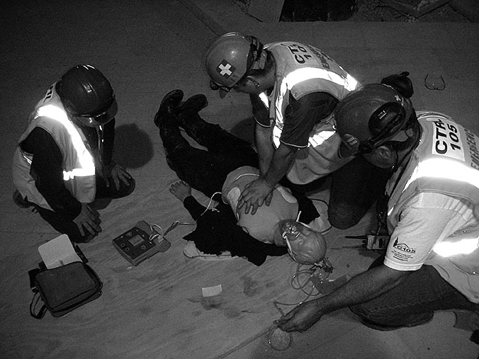
Are we performing resuscitation for long enough? In this study the authors wanted to know for in-hospital cardiac arrests, how long resuscitation attempts should be continued before termination of efforts. While this is an in-hospital study, the answer could be relevant for pre-hospital guidelines. The authors investigated whether duration of resuscitation attempts varied between hospitals and whether patients at hospitals that attempt resuscitation for longer have higher survival rates than do those at hospitals with shorter durations of resuscitation efforts.

Between 2000 and 2008, 64,339 patients with cardiac arrests were identified at 435 US hospitals within the Get with the Guidelines – Resuscitation registry. For each hospital, the median duration of resuscitation before termination of efforts in non-survivors was. Multilevel regression models were used to assess the association between the length of resuscitation attempts and risk-adjusted survival. Primary endpoints were immediate survival with return of spontaneous circulation (ROSC) during cardiac arrest and survival to hospital discharge.

31,198 of 64,339 (48·5%) patients achieved return of spontaneous circulation and 9,912 (15·4%) survived to discharge. For patients achieving return of spontaneous circulation, the median duration of resuscitation was 12 min compared with 20 min for non-survivors. Compared with patients at hospitals in the quartile with the shortest median resuscitation attempts in non-survivors (16 min), those at hospitals in the quartile with the longest attempts (25 min) had a higher likelihood of return of spontaneous circulation and survival to discharge.

It was found that duration of resuscitation attempts varies between hospitals. The authors believe that although they cannot define an optimum duration for resuscitation attempts on the basis of these observational data, their findings suggest that efforts to systematically increase the duration of resuscitation could improve survival in this high-risk population.

According to the Australian Resuscitation Council, there is substantial variability in the approaches to termination of resuscitation attempts during cardiac arrest management and factors taken into account include whether the arrest was witnessed, whether bystander CPR was provided, the duration of cardiac arrest, and associated co-morbidities. Just looking to the US for examples of their termination of resuscitation recommendations, while advanced level providers have a minimum time of 20 minutes specified, basic level providers can terminate after 6 minutes (3 full rounds) of CPR and AED analysis.

A large proportion of pre-hospital guidelines specify that resuscitation cannot be terminated until after 20 minutes of ALS care, which once you include the initial CPR, defibrillation, venous access and airway management skills would probably amount to a resuscitation time of 25 minutes or longer which would follow the study’s findings of a greater likelihood of survival and ROSC. However, where there are no guidelines specifying time limits, or those that follow the ‘no ROSC within 3 full cycles of CPR and AED analysis’ rule, should be we saying that these ambulance crews should be staying on scene for longer (maybe even 20 – 25 minutes), especially when considering the poor effectiveness of CPR during extrication and in a moving ambulance.

http://www.thelancet.com/journals/lancet/article/PIIS0140-6736(12)60862-9/abstract

While on the subject of poor effectiveness of CPR, a recent study has found that fatigue can affect chest compression delivery within the second minute of CPR, providing more evidence of the importance of, wherever possible, changing the chest compression provider after every rhythm analysis..

Rescuer fatigue under the 2010 ERC guidelines and its effect on cardiopulmonary resuscitation (CPR) performance

Emerg Med J 2012 Published online

This study sought to determine the impact of the 2010 resuscitation guidelines on rescuer fatigue and cardiopulmonary resuscitation (CPR) performance.

62 Health science students performed 5 min of conventional CPR in accordance with the 2010 ERC guidelines. A SkillReporter manikin was used to objectively assess temporal change in determinants of CPR quality. Participants subjectively reported their end-fatigue levels, using a visual analogue scale, and the point at which they believed fatigue was affecting CPR delivery.

49 (79%) participants reported that fatigue affected their CPR performance, at an average of 167 s. End fatigue averaged 49.5/100. The proportion of chest compressions delivered correctly decreased from 52% in min 1 to 39% in min 5, approaching significance. A significant decline in chest compressions reaching the recommended depth occurred between the first (53%) and fifth (38%) min. Almost half this decline (6%) was between the first and second minutes of CPR. Neither chest compression rate, nor rescue breath volume, were affected by rescuer fatigue.

Fatigue affects chest compression delivery within the second minute of CPR under the 2010 ERC guidelines, according to the authors, and is poorly judged by rescuers. Rescuers should, therefore, be encouraged to interchange after 2 min of CPR delivery. Team leaders should be advised to not rely on rescuers to self-report fatigue, and should, instead, monitor for its effects.

http://emj.bmj.com/content/early/2012/07/31/emermed-2012-201610.abstract

Endotracheal intubation versus supraglottic airway insertion in out-of-hospital cardiac arrest

Resuscitation, Volume 83, Issue 9, September 2012, Pages 1047–1048

To simplify airway management and minimise interruptions to cardiopulmonary resuscitation (CPR) chest compression, there has been an increase in the use of supraglottic airway (SGA) devices instead of endotracheal intubation (ETI) as the primary airway adjunct in out-of-hospital cardiac arrest (OHCA). For this research the authors compared the outcomes of patients receiving ETI with those receiving SGA following OHCA.

A secondary analysis of data from the multicenter Resuscitation Outcomes Consortium (ROC) PRIMED trial was undertaken with adult non-traumatic OHCA receiving successful SGA insertion (King Laryngeal Tube, Combitube, and Laryngeal Mask Airway) or successful ETI studied. The primary outcome was survival to hospital discharge with satisfactory functional status (Modified Rankin Scale ≤3). Secondary outcomes included return of spontaneous circulation (ROSC), 24-h survival, major airway or pulmonary complications (pulmonary oedema, internal thoracic or abdominal injuries, acute lung injury, sepsis, and pneumonia). Using multivariable logistic regression, the authors studied the association between out-of-hospital airway management method (ETI vs. SGA) and OHCA outcomes, adjusting for confounders.

Of 10,455 adult OHCA, 8487 (81.2%) received ETI and 1968 (18.8%) received SGA. Survival to hospital discharge with satisfactory functional status was4.7% for ETI, compared with 3.9% for SGA. Compared with successful SGA, successful ETI was associated with increased survival to hospital discharge, ROSC and 24-h survival. ETI was not associated with secondary airway or pulmonary.

In this secondary analysis of data from the trial, ETI was associated with improved outcomes when compared with SGA insertion after OHCA.

Following the various published research studies which states that pre-hospital tracheal intubation can be associated with no benefit or is harmful this article could be welcomed by some as good news, providing the more evidence for they importance of paramedic intubation, but it may not be as simple as that.

The clinical trial was never intended to be used for the evaluation of airway management techniques and did not record such variables as time of airway insertion (for example before or after ROSC), number of airway attempts, whether the airway was displaced during resuscitation or transportation and interruptions to chest compressions. Likewise, the decision to intubate over the use of a SGA was left to the decision of the attending paramedic (no randomised trial) and, as the SGA is still thought to by some as a failed intubation device, could be the reason that a SGA was used, without intubation being attempted, in only 12.2% of cases. While taking this into consideration, this is a trial involving a large number of patients – over 10,000 – and the authors might have found some evidence in favour for the, often argued subject of whether paramedics should intubate.

It is clear that more research, not only randomised comparisons on airway devices with minimal variables (yes, I know how hard this is in the cardiac arrest situation but large number studies may reduce the effect of such variables) is required, but also a comparison on the different types of SGA device. As SGAs used to be considered as a failed intubation tool, the disadvantages were sometimes accepted, however with the increased use, or especially when considered as a primary adjunct, the risks associated with a SGA should be considered. In animal models it has been shown that there has been 15–50% decrease in carotid blood flow observed with the use of SGA (King LT, LMA and Combitube) compared with ETI, thought to be caused by the cuff and/or balloons applying pressure on the carotid artery. If this is found to be true in humans, this could affect neurological outcome and survival.  Other reported complications associated with Combitube use including aspiration pneumonitis, airway and esophageal injuries, and cranial nerve injury, similar adverse events may be possible with King LT or LMA use in OHCA.

One study which should provide interesting reading is the randomised trial of the I-gel versus LMA Supreme versus standard care in OHCA currently being undertaken within the UK (http://www.controlled-trials.com/ISRCTN18528625/18528625), especially as the I-gel does not have an inflatable cuff or balloon which not only decreases the time of insertion but could also reduce the pressure on the carotid artery when compared with other SGA devices.

Airway management is only a part of the resuscitation attempt, studies face a huge number of variables which could affect survival and ROSC, for example, the time taken until CPR started and first shock, the training and experience levels of paramedics, any difficult extrication causing prolonged interruptions to chest compressions, hyperventilation (both during CPR and post ROSC) and the use of therapeutic hypothermia. Until we have more evidence which clearly identifies the most appropriate airway management technique, I believe we should continue doing what we already know that improves ROSC and survival, for example:

  • Early recognition of cardiac arrest and ambulance activation;
  • Early CPR by bystanders and community responders;
  • Minimise time to first shock through the use of public access defibrillators, community responders and rapid ambulance response;
  • Good quality CPR with minimal interruptions and frequent changes to CPR providers to prevent tiring, this may also include the use of mechanical CPR devices but more research on these would be nice;
  • Utilisation of appropriate airway management devices without causing interruptions to compressions, this may be either an ETI or SGA, but the important thing is that chest compressions are not interrupted and hypoxia minimised during airway management attempts. Where resources are limited, it may be appropriate for a SGA device to be inserted very early while chest compressions are being undertaken and then when/ if ROSC is obtained, changed to a ET tube for the transportation phase and ROSC management strategy; and
  • Use of adrenaline, yes, the jury is still out on this but studies have shown that it can increase the likelihood of ROSC. Once we have ROSC, strategies such as therapeutic hypothermia, maintenance of blood pressure and arrhythmias, adequate ventilation and oxygenation (including the use of end-tidal capnography) and transport to PCI capable centres, where appropriate, should become our focus.

http://www.sciencedirect.com/science/article/pii/S0300957212002705